Publications

Density Fitting, Fast & Gentle

Published online 31 July 2023 in PLOS Computational Biology (doi 10.1371/journal.pcbi.1011255):

Gentle and fast all-atom model refinement to cryo-EM densities via a maximum likelihood approach

Christian Blau, Linnea YvonnesdotterErik Lindahl

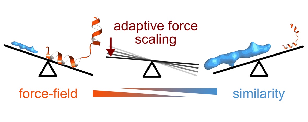
Better detectors and automated data collection have generated a flood of high-resolution cryo-EM maps, which in turn has renewed interest in improving methods for determining structure models corresponding to these maps. However, automatically fitting atoms to densities becomes difficult as their resolution increases and the refinement potential has a vast number of local minima. In practice, the problem becomes even more complex when one also wants to achieve a balance between a good fit of atom positions to the map, while also establishing good stereochemistry or allowing protein secondary structure to change during fitting. Here, we present a solution to this challenge using a maximum likelihood approach by formulating the problem as identifying the structure most likely to have produced the observed density map. This allows us to derive new types of smooth refinement potential—based on relative entropy—in combination with a novel adaptive force scaling algorithm to allow balancing of force-field and density-based potentials. In a low-noise scenario, as expected from modern cryo-EM data, the relative-entropy based refinement potential outperforms alternatives, and the adaptive force scaling appears to aid all existing refinement potentials. The method is available as a component in the GROMACS molecular simulation toolkit.

Read the full publication here.

Publications

MD-Fitting a Membrane Protein

Published 11 July 2023 in Biophysical Journal (doi 10.1016/j.bpj.2023.05.033):

Automated simulation-based membrane protein refinement into cryo-EM data

Linnea Yvonnesdotter, Urška Rovšnik, Christian Blau, Marie Lycksell, Rebecca J Howard, Erik Lindahl

The resolution revolution has increasingly enabled single-particle cryogenic electron microscopy (cryo-EM) reconstructions of previously inaccessible systems, including membrane proteins—a category that constitutes a disproportionate share of drug targets. We present a protocol for using density-guided molecular dynamics simulations to automatically refine atomistic models into membrane protein cryo-EM maps. Using adaptive force density-guided simulations as implemented in the GROMACS molecular dynamics package, we show how automated model refinement of a membrane protein is achieved without the need to manually tune the fitting force ad hoc. We also present selection criteria to choose the best-fit model that balances stereochemistry and goodness of fit. The proposed protocol was used to refine models into a new cryo-EM density of the membrane protein maltoporin, either in a lipid bilayer or detergent micelle, and we found that results do not substantially differ from fitting in solution. Fitted structures satisfied classical model-quality metrics and improved the quality and the model-to-map correlation of the x-ray starting structure. Additionally, the density-guided fitting in combination with generalized orientation-dependent all-atom potential was used to correct the pixel-size estimation of the experimental cryo-EM density map. This work demonstrates the applicability of a straightforward automated approach to fitting membrane protein cryo-EM densities. Such computational approaches promise to facilitate rapid refinement of proteins under different conditions or with various ligands present, including targets in the highly relevant superfamily of membrane proteins.

Read the full publication here.

Publications

Ivermectin by NMR

Published 23 February 2023 in ACS Chemical Neuroscience (doi 10.1021/acschemneuro.2c00783):

Structural elucidation of ivermectin binding to α7nAChR and the induced channel desensitization

Vasyl Bondarenko, Qiang Chen, Kevin Singewald, Nandan Haloi, Tommy S. Tillman, Rebecca J HowardErik Lindahl, Yan Xu, Pei Tang

The α7 nicotinic acetylcholine receptor (α7nAChR) mediates signaling in the central nervous system and cholinergic anti-inflammatory pathways. Ivermectin is a positive allosteric modulator of a full-length α7nAChR and an agonist of the α7nAChR construct containing transmembrane (TMD) and intracellular (ICD) domains, but structural insights of the binding have not previously been determined. Here, combining nuclear magnetic resonance as a primary experimental tool with Rosetta comparative modeling and molecular dynamics simulations, we have revealed details of ivermectin binding to the α7nAChR TMD + ICD and corresponding structural changes in an ivermectin-induced desensitized state. Ivermectin binding was stabilized predominantly by hydrophobic interactions from interfacial residues between adjacent subunits near the extracellular end of the TMD, where the inter-subunit gap was substantially expanded in comparison to the apo structure. The ion-permeation pathway showed a profile distinctly different from the resting-state profile but similar to profiles of desensitized α7nAChR. The ICD also exhibited structural changes, including reorientation of the MX and h3 helices relative to the channel axis. The resulting structures of the α7nAChR TMD + ICD in complex with ivermectin provide opportunities for discovering new modulators of therapeutic potential and exploring the structural basis of cytoplasmic signaling under different α7nAChR functional states.

Read the full publication here.

Publications

Pi in the Gating Cycle of NavAb

Published 14 December 2022 in Journal of General Physiology (doi 10.1085/jgp.202213214):

An α–π transition in S6 shapes the conformational cycle of the bacterial sodium channel NavAb

Koushik Choudhury, Rebecca J Howard, Lucie Delemotte

Voltage-gated sodium channels play an important role in electrical signaling in excitable cells. In response to changes in membrane potential, they cycle between nonconducting and conducting conformations. With recent advances in structural biology, structures of sodium channels have been captured in several distinct conformations, which are thought to represent different functional states. However, it has been difficult to capture the intrinsically transient open state. We recently showed that a proposed open state of the bacterial sodium channel NavMs was not conductive and that a conformational change involving a transition to a π-helix in the pore-lining S6 helix converted this structure into a conducting state. However, the relevance of this structural feature in other sodium channels, and its implications for the broader gating cycle, remained unclear. Here, we propose a comparable open state of another class of bacterial channel from Aliarcobacter butzleri (NavAb) with characteristic pore hydration, ion permeation, and drug binding properties. Furthermore, we show that a π-helix transition can lead to pore opening and that such a conformational change blocks fenestrations in the inner helix bundle. We also discover that a region in the C-terminal domain can undergo a disordering transition proposed to be important for pore opening. These results support a role for a π-helix transition in the opening of NavAb, enabling new proposals for the structural annotation and drug modulation mechanisms in this important sodium channel model.

Read the full publication here.

Publications

Divalents and Dynamic Domains

Published 8 December 2022 in Proceedings of the National Academy of Sciences of the USA (doi 10.1073/pnas.2210669119):

Biophysical characterization of calcium-binding and modulatory-domain dynamics in a pentameric ligand-gated ion channel

Marie Lycksell, Urška Rovšnik, Anton Hanke, Anne Martel, Rebecca J Howard, Erik Lindahl

Pentameric ligand-gated ion channels (pLGICs) perform electrochemical signal transduction in organisms ranging from bacteria to humans. Among the prokaryotic pLGICs, there is architectural diversity involving N-terminal domains (NTDs) not found in eukaryotic relatives, exemplified by the calcium-sensitive channel (DeCLIC) from a Desulfofustis deltaproteobacterium, which has an NTD in addition to the canonical pLGIC structure. Here, we have characterized the structure and dynamics of DeCLIC through cryoelectron microscopy (cryo-EM), small-angle neutron scattering (SANS), and molecular dynamics (MD) simulations. In the presence and absence of calcium, cryo-EM yielded structures with alternative conformations of the calcium-binding site. SANS profiles further revealed conformational diversity at room temperature beyond that observed in static structures, shown through MD to be largely attributable to rigid-body motions of the NTD relative to the protein core, with expanded and asymmetric conformations improving the fit of the SANS data. This work reveals the range of motion available to the DeCLIC NTD and calcium-binding site, expanding the conformational landscape of the pLGIC family. Further, these findings demonstrate the power of combining low-resolution scattering, high-resolution structural, and MD simulation data to elucidate interfacial interactions that are highly conserved in the pLGIC family.

Read the full publication here.

Publications

Lipid Sites in nAChR States

Published 17 October 2022 in Proceedings of the National Academy of Sciences of the USA (doi 10.1073/pnas.2208081119):

Differential interactions of resting, activated, and desensitized states of the α7 nicotinic acetylcholine receptor with lipidic modulators

Yuxuan Zhuang, Colleen M Noviello, Ryan E Hibbs, Rebecca J Howard, Erik Lindahl

The α7 nicotinic acetylcholine receptor is a pentameric ligand-gated ion channel that modulates neuronal excitability, largely by allowing Ca2+ permeation. Agonist binding promotes transition from a resting state to an activated state, and then rapidly to a desensitized state. Recently, cryogenic electron microscopy (cryo-EM) structures of the human α7 receptor in nanodiscs were reported in multiple conformations. These were selectively stabilized by inhibitory, activating, or potentiating compounds. However, the functional annotation of these structures and their differential interactions with unresolved lipids and ligands remain incomplete. Here, we characterized their ion permeation, membrane interactions, and ligand binding using computational electrophysiology, free-energy calculations, and coarse-grained molecular dynamics. In contrast to nonconductive structures in apparent resting and desensitized states, the structure determined in the presence of the potentiator PNU-120596 was consistent with an activated state permeable to Ca2+. Transition to this state was associated with compression and rearrangement of the membrane, particularly in the vicinity of the peripheral MX helix. An intersubunit transmembrane site was implicated in selective binding of either PNU-120596 in the activated state or cholesterol in the desensitized state. This substantiates functional assignment of all three lipid-embedded α7-receptor structures with ion-permeation simulations. It also proposes testable models of their state-dependent interactions with lipophilic ligands, including a mechanism for allosteric modulation at the transmembrane subunit interface.

Read the full publication here.

Publications

Modeling Bimodal Modulation

Published 6 August 2022 in Nature Communications (doi 10.1038/s41467-022-32212-4):

Structural and dynamic mechanisms of GABAA receptor modulators with opposing activities

Shaotong Zhu, Akshay Sridhar, Jinfeng Teng, Rebecca J HowardErik Lindahl & Ryan E Hibbs

γ-Aminobutyric acid type A (GABAA) receptors are pentameric ligand-gated ion channels abundant in the central nervous system and are prolific drug targets for treating anxiety, sleep disorders and epilepsy. Diverse small molecules exert a spectrum of effects on γ-aminobutyric acid type A (GABAA) receptors by acting at the classical benzodiazepine site. They can potentiate the response to GABA, attenuate channel activity, or counteract modulation by other ligands. Structural mechanisms underlying the actions of these drugs are not fully understood. Here we present two high-resolution structures of GABAA receptors in complex with zolpidem, a positive allosteric modulator and heavily prescribed hypnotic, and DMCM, a negative allosteric modulator with convulsant and anxiogenic properties. These two drugs share the extracellular benzodiazepine site at the α/γ subunit interface and two transmembrane sites at β/α interfaces. Structural analyses reveal a basis for the subtype selectivity of zolpidem that underlies its clinical success. Molecular dynamics simulations provide insight into how DMCM switches from a negative to a positive modulator as a function of binding site occupancy. Together, these findings expand our understanding of how GABAA receptor allosteric modulators acting through a common site can have diverging activities.

Read the full publication here.

Publications

COVID-E Biophysics

Published 1 October 2022 in Biochimica et Biophysica Acta – Biomembranes (doi 10.1016/j.bbamem.2022.183994):

Probing effects of the SARS-CoV-2 E protein on membrane curvature and intracellular calcium

Aujan Mehregan, Sergio Pérez-Conesa, Yuxuan Zhuang, Ahmad Elbahnsi, Diletta Pasini, Erik Lindahl, Rebecca J Howard, Chris Ulens, Lucie Delemotte

SARS-CoV-2 contains four structural proteins in its genome. These proteins aid in the assembly and budding of new virions at the ER-Golgi intermediate compartment (ERGIC). Current fundamental research efforts largely focus on one of these proteins – the spike (S) protein. Since successful antiviral therapies are likely to target multiple viral components, there is considerable interest in understanding the biophysical role of its other structural proteins, in particular structural membrane proteins. Here, we have focused our efforts on the characterization of the full-length envelope (E) protein from SARS-CoV-2, combining experimental and computational approaches. Recombinant expression of the full-length E protein from SARS-CoV-2 reveals that this membrane protein is capable of independent multimerization, possibly as a tetrameric or smaller species. Fluorescence microscopy shows that the protein localizes intracellularly, and coarse-grained MD simulations indicate it causes bending of the surrounding lipid bilayer, corroborating a potential role for the E protein in viral budding. Although we did not find robust electrophysiological evidence of ion-channel activity, cells transfected with the E protein exhibited reduced intracellular Ca2+, which may further promote viral replication. However, our atomistic MD simulations revealed that previous NMR structures are relatively unstable, and result in models incapable of ion conduction. Our study highlights the importance of using high-resolution structural data obtained from a full-length protein to gain detailed molecular insights, and eventually permitting virtual drug screening.

Read the full publication here.

Publications

Pi-Bulge to Open

Published 4 January 2022 in Biophysical Journal (doi 10.1016/j.bpj.2021.12.010):

An open state of a voltage-gated sodium channel involving a π-helix and conserved pore-facing asparagine

Koushik Choudhury, Marina A Kasimova, Sarah McComas, Rebecca J Howard, Lucie Delemotte

Voltage-gated sodium (Nav) channels play critical roles in propagating action potentials and otherwise manipulating ionic gradients in excitable cells. These channels open in response to membrane depolarization, selectively permeating sodium ions until rapidly inactivating. Structural characterization of the gating cycle in this channel family has proved challenging, particularly due to the transient nature of the open state. A structure from the bacterium Magnetococcus marinus Nav (NavMs) was initially proposed to be open, based on its pore diameter and voltage-sensor conformation. However, the functional annotation of this model, and the structural details of the open state, remain disputed. In this work, we used molecular modeling and simulations to test possible open-state models of NavMs. The full-length experimental structure, termed here the α-model, was consistently dehydrated at the activation gate, indicating an inability to conduct ions. Based on a spontaneous transition observed in extended simulations, and sequence/structure comparison to other Nav channels, we built an alternative π-model featuring a helix transition and the rotation of a conserved asparagine residue into the activation gate. Pore hydration, ion permeation, and state-dependent drug binding in this model were consistent with an open functional state. This work thus offers both a functional annotation of the full-length NavMs structure and a detailed model for a stable Nav open state, with potential conservation in diverse ion-channel families.

Read the full publication here.

Publications

From Leak to Disease

Published December 2021 in Journal of Biological Chemistry (doi 10.1016/j.jbc.2021.101355):

A missense mutation converts the Na+,K+-ATPase into an ion channel and causes therapy-resistant epilepsy

Sofia Ygberg, Evgeny E Akkuratov*, Rebecca J Howard*, Fulya Taylan, Daniel C Jans*, Dhani R Mahato, Adriana Katz, Paula F Kinoshita, Benjamin Portal, Inger Nennesmo, Maria Lindskog, Steven JD Karlish, Magnus Andersson*, Anna Lindstrand, Hjalmar Brismar*, Anita Aperia*

*Current and former members of the SciLifeLab biophysics community

The ion pump Na+,K+-ATPase is a critical determinant of neuronal excitability; however, its role in the etiology of diseases of the central nervous system (CNS) is largely unknown. We describe here the molecular phenotype of a Trp931Arg mutation of the Na+,K+-ATPase catalytic α1 subunit in an infant diagnosed with therapy-resistant lethal epilepsy. In addition to the pathological CNS phenotype, we also detected renal wasting of Mg2+. We found that membrane expression of the mutant α1 protein was low, and ion pumping activity was lost. Arginine insertion into membrane proteins can generate water-filled pores in the plasma membrane, and our molecular dynamic (MD) simulations of the principle states of Na+,K+-ATPase transport demonstrated massive water inflow into mutant α1 and destabilization of the ion-binding sites. MD simulations also indicated that a water pathway was created between the mutant arginine residue and the cytoplasm, and analysis of oocytes expressing mutant α1 detected a nonspecific cation current. Finally, neurons expressing mutant α1 were observed to be depolarized compared with neurons expressing wild-type protein, compatible with a lowered threshold for epileptic seizures. The results imply that Na+,K+-ATPase should be considered a neuronal locus minoris resistentia in diseases associated with epilepsy and with loss of plasma membrane integrity.

Read the full publication here.